Introduction
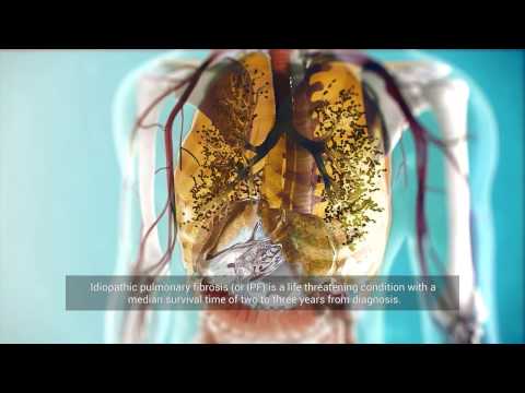
Pulmonary fibrosis is a disease in which there is a pathological formation of scar tissue in the lungs so that they cannot fully perform their function to provide the body with oxygen. This condition significantly worsens the quality of life of the patient. In some cases, the progression of the disease may lead to lung and/or heart failure and death. Modern medical drugs can’t reverse the pathological changes in lung fibrosis. Alternative methods of treating pathology can include stem cells, since their studied properties involve the ability to stop the inflammatory process, as well as to stimulate the recovery of damaged tissues.
About Pulmonary Fibrosis
Pulmonary fibrosis is associated with lung tissue scarring due to its fibrotic degeneration. The growth of scar tissue, caused as a rule by inflammation, leads to compression of the alveoli, which can no longer fully perform their function, which is to ensure gas exchange in the body. Alveoli are the pulmonary vesicles in which inhaled air contacts the blood. Fibrotic lesions can affect a part of the lung or the entire organ, in one or both lungs.
Pulmonary fibrosis, or pneumofibrosis, is a progressive illness with irreversible changes in lung tissue, whose complications and consequences include:
- Chronic respiratory failure (insufficient oxygen to the body).
- Pulmonary hypertension (high blood pressure in the pulmonary arteries).
- Chronic pulmonary heart and other cardiovascular diseases.
- Lung cancer.
- Secondary respiratory infection (including the development of pneumonia with a high risk of death).
The Causes of Pulmonary Fibrosis
It is not always possible to determine the exact reason that causes lung scarring. Pathological growth of connective tissue in the lungs may be provoked by allergies, lung infections, genetic factors, radiation, trauma and other processes and circumstances.
When it is impossible to determine the exact cause of the pathology, we are talking about idiopathic pulmonary fibrosis (IPF). In such a disease, a combination of environmental exposure, genetic and other unidentified factors is considered a provocateur.
There is also evidence that smoking (both in the present and the past), plus a burdened family history (noted in up to 20% of patients), plays a role. When at least two or more primary biological family members (parent, sibling, or child) have had clinical manifestations of IPF, the disease is classified as familial interstitial pneumonia (FIP). First-degree relatives of patients with FIP have a greatly increased risk of developing interstitial lung disease (ILD).
Interstitial Pulmonary Fibrosis
Interstitial lung disease (ILD), also known as diffuse parenchymal lung disease (DPLD), is a group of pathologies, including pulmonary fibrosis, in which the interstitial (connective tissue of the lung) is damaged. ILD is associated with inflammation and scarring of the lung tissue.
Interstitial tissue is the connective tissue of the lungs that serves as the backbone for the alveoli and blood vessels. In fibrosis, due to the excessive growth of connective tissue in the lung, alveoli are compressed and they are subsequently replaced with connective tissue. As a result, gas exchange in the alveoli becomes difficult, resulting in hypoxia – the body begins to suffer from a lack of oxygen.
Unlike idiopathic pulmonary fibrosis (IPF), the cause of ILD can be reliably determined. These reasons include:
- infectious lesions;
- autoimmune / connective tissue diseases;
- genetic predisposition;
- malignancies;
- harmful inhaled substances (mercury vapour, mineral dust, etc);
- certain medications (for example, some antiarrhythmic drugs, nitrofurantoin and others).
Contact us
Get a free online consultation to learn about the expected results of stem cell therapy, its cost and duration.
The coronavirus pandemic has increased the number of all cases of lung fibrosis. Now it is known that lung scarring is one of the most frequent and most serious consequences of viral exposure on the human body. The mechanism of development of pulmonary fibrosis caused by COVID-19 is associated with an incorrect immune response, which leads to damage of lung epithelial cells and microvascular endothelial cells, especially in combination with artificial lung ventilation.
Moreover, according to researchers and statistical data, it becomes clear that patients with mild COVID-19 infection, even after improvement and discharge from the hospital, have a potential hidden risk of developing pulmonary fibrosis.
To prevent the progression of pulmonary fibrosis, induced by the coronavirus, it is essential to start the treatment of this complication as early as possible.
Signs & Symptoms of Pulmonary Fibrosis
The clinical course of pulmonary fibrosis is associated with a decrease in lung volume, which forces the body to adapt to this negative consequence. Compensatory changes manifest as shortness of breath, which is the most frequent initial symptom reported by patients with IPF. At first, shortness of breath occurs only during physical exertion but later – also at rest.
The patient with IPF also faces symptoms such as a dry cough, fatigue and unexplained weight loss.
Also, clinical features of an underlying connective tissue disease (CTD), such as arthralgia (aching in several joints) or sicca symptoms (dryness of the exocrine glands, particularly the eyes and mouth), might be observed.
The main external sign of IPF is known as ‘clubbed fingers’, which is a deformity of the fingers or toes characterized by enlargement of the tips of the digits. This symptom is associated with chronically low blood levels of oxygen.
The initial stages of the disease most often occur without manifestations. For example, in asymptomatic individuals at risk for familial interstitial pneumonia (FIP), signs of histologic changes in the lung tissue can be diagnostically identified.
Diagnosing Pulmonary Fibrosis
There is an opinion that lung fibrosis as a disease is insufficiently diagnosed. Most often, it is detected at a fairly late stage. Commonly, the disease is identified by an abnormality on a high-resolution computed tomography (HRCT) scan of the chest.
There is evidence that chest radiography is less useful than HRCT in evaluating patients with suspected IPF.
Bronchoscopy with bronchoalveolar lavage (BAL) and transbronchial biopsy are also used to determine the condition of lung tissue.
Also, laboratory diagnostics of various indicators (biological markers) can provide objectively measurable data about processes occurring in the body – both normal and pathological. In pulmonary fibrosis, these biomarkers help to diagnose the disease, reveal the disease susceptibility and predict specific drug efficacy. They can also accurately predict the course of the disease and its prognosis in a patient with IPF.
Traditional and Modern Treatments for ILD & Lung Fibrosis
While pulmonary fibrosis may take years to develop, most patients with the disease are diagnosed in its advanced stage, when little can be done to influence its progression. The existing treatments are aimed at maintaining the patient’s lung performance and an acceptable standard of living.
In the common method of treatment of pneumofibrosis, adrenal cortex hormones (prednisone) are used, and if they are not effective enough, azathioprine and cyclophosphane are used. Also, assigned to oxygen for inhalation. Recently, medications pirfenidone and nintedanib have been shown to slow IPF progression.
However, pulmonary fibrosis is a progressive disease and no antifibrotic agents or treatment (short of a lung transplantation) are known to impact the poor survival rate associated with this disease.
Alternative methods of treating lung fibrosis and ILD include the use of stem cell products. Pathological changes occurring in the lungs with fibrosis are considered irreversible, due to the fact that the body has a poor ability to heal scar tissue. However, when we launch stem cells into the body in the amount of a therapeutic dose, this is often enough to start the regeneration processes and slow down the course of scarring of the lung tissue. These cells can be injected either through an IV drip (intravenously) or by inhalation for targeted delivery of cells to the damaged area.
Get a free online consultation
Contact us to learn what benefits you can get from stem cell therapy.
Stem Cell Therapy for Pulmonary Fibrosis
Cell-based therapy is known as a modern experimental treatment that uses the body’s ability to treat itself.
How do infused stem cells act in the fibrosis? The peculiarity of the ‘work’ of stem cells in the body is that they respond to signals sent by cells of damaged tissues, and produce cytokines (protein molecules that transmit intercellular signals) in response, which stimulate the formation of the new required cells.
Studies of allogeneic (donor) mesenchymal stem cells (MSCs) have shown that they hone in to sites of lung injury and contribute to tissue regeneration, decreasing chronic airway inflammation and restoration of alveolar fluid balance in acute lung injury.
Currently, further stem cell clinical trials for lung disease are being conducted.
What Are the Possible Results After Stem Cell Therapy?
Using stem cells for intravenous infusions in the treatment of lung fibrosis, we can achieve the following effects:
- Slowing down or halting the scarring process, by ‘calming’ the patient’s immune system (stem cells have an anti-inflammatory ability).
- Partial regeneration of lung tissue.
- Formation of new vascular networks, which contribute to tissue renewal.
- Relief of the patient’s symptoms.
According to a clinical trial, endobronchial infusions of stem cells increased the number of indicators for quality of life. 86% of the participants of the study, who were observed for 12 months, demonstrated stable functional status and physical performance.
Thus, the main role of therapy, based on the stem cell introduction, is slowing down the progression of the disease, stabilising lung function and maintaining the current state of the patient.
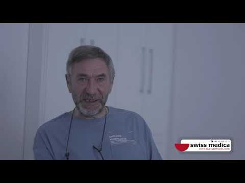
Watch the video review of the patients with COPD (in Italian) to learn how stem cell therapy can help in lung disease:
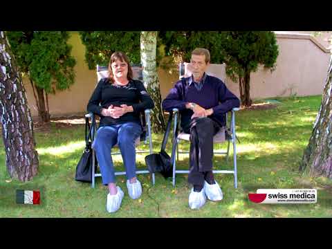
One more testimonial:
Safety of Stem Cells in Pulmonary Fibrosis
Сlinical trials of therapy, based on adult stem cells, demonstrated an acceptable safety profile of this approach. In particular, it was reported that during the entire period of a study using 72 infusions, no side effects were reported that could be considered serious or clinically significant [19].
It is currently being debated whether MSCs can differentiate into fibroblasts, given their common mesodermal origin, and accelerate the fibrous cascade. However, there are no preclinical or human studies that have demonstrated this relationship.
None of the patients experienced any abnormal tissue formation, as reported by a whole-body CT scan performed 12 months after the first stem cell administration. The same positive safety result was obtained 24 months after the first infusion.
Another study demonstrates that infusions of stem cells were well-tolerated in all patients and no treatment-emergent serious adverse events were reported.
Contact our medical advisor
Contact us to learn about the expected results of the treatment, its cost and duration.
List of References
World Health Organisation. The top 10 causes of death. 24 May 2018.
Mesenchymal Stem Cell as Therapeutic Modality in Interstitial Pulmonary Fibrosis. Clinical Trial.